Basic Health
A “healthy smile” does not mean a mouth full of perfectly arranged, bleach-white teeth. It means a mouth that is free of diseases such as caries, periodontitis, oral cancer, viral manifestations, chronic ulcers, autoimmune disease and much, much more. Many of the signs that a dentist diagnoses in the mouth can be warning signs or causes of systemic illness. Think of the mouth as a window to the rest of the body.

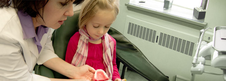
At each dental check-up, a full intra-oral and head/neck exam is done to ensure that your oral health is properly monitored and that you receive the information you need to make important health decisions. The type and frequency of radiographs will be determined by your personal risk factors. A periodontal charting will be completed at the first visit and updated periodically.
DENTAL CARIES
Caries is the name for the disease that causes “cavities” in teeth. It is classified as a disease because it is caused by bacteria that damage the body. These bacteria feed on carbohydrates in the mouth, converting them to acid that erodes the hard enamel shell of the teeth. Once this barrier is breached, the bacteria move further into the tooth, growing in numbers and eroding more and more tooth structure. Untreated, this infection eventually makes it to the nerve of the tooth, causing pain, and ultimately killing the nerve, blood vessels and other vital cells inside the tooth. This may cause a dark discoloration of the tooth, but far worse, the infection can then spread down the root of the tooth, into the bone and frequently into the soft tissue of the head and/or neck.
A tooth infection that causes swelling of the face or neck must be treated very seriously and requires immediate dental and/or medical emergency treatment.
Fortunately, caries need not become a medical emergency. The purpose of regular dental care is primarily prevention. Each person is an individual in terms of their risk for caries. This risk is determined by factors such as existing decay, oral hygiene, diet, anatomy and salivary flow rate. Your individual risk is assessed by your dental hygienist and dentist, and based on this, recommendations will be made to help you become and stay caries-free.
Caries on the biting surfaces or outer surfaces of the teeth may be detected visually or by “feel” with a dental instrument. However, the more common location for dental caries is in between teeth. To see caries between teeth, a dentist uses a radiograph, which is a picture of the teeth made by an X-ray. Radiographs are very helpful for finding caries early when the decay is very small and can be simply, painlessly and economically fixed with a small filling.
PERIODONTAL DISEASE
Periodontal Disease begins as gingivitis when a film of bacteria called plaque builds up on teeth around the gum line causing redness, swelling or bleeding of the gums.
Left untreated, however, the plaque becomes calcified and turns into calculus (think “barnacles”) on the root surfaces of the teeth, which cannot be removed at home. Calculus creates a nice environment for the attachment of more bacteria and over time, this infection causes damage to the gingiva, “gum recession”. But much worse, the interaction between the inflamed gingiva, and the bacteria causes periodontal disease, the disintegration of the ligament and bone that holds the teeth into the jawbone. Without proper treatment, the teeth begin to loosen and are eventually lost. Periodontal disease is the most common cause for tooth loss (more than cavities!), and is often overlooked by patients simply because it is usually not painful at all.
Periodontal disease is unfortunately not a disease that affects only the mouth. Medical research over the past 20 years has found many links between periodontal disease and systemic disease. Because periodontal disease causes inflammation, the gums bleed very easily. Every time capillaries in the gums open up and bleed, there is the potential to carry the organisms from the infected gums into distant sites of the body. Periodontitis sets up a chronic inflammatory state that the body must support. Research has demonstrated an association between periodontal disease and other chronic inflammatory conditions, such as diabetes, cardiovascular disease, stroke and Alzheimer’s disease. Periodontitis carries an increased risk for patients with existing COPD, and has been associated with a markedly higher risk for pancreatic cancer. Therefore, treatment and prevention of periodontal disease not only establishes a healthy mouth but may also provide risk reduction and management of other systemic conditions.
ORAL CANCER
The incidence of oral-pharyngeal cancer continues to be on the rise, topping 36,000 new cases in 2011. There is currently only a 57% 5-year survival rate, much worse than that of other cancers such as cervical, laryngeal, testicular, thyroid, and skin.
One of the reasons for such a poor survival rate is that many of these cases are not detected earlier. This makes it critically important for routine oral cancer screenings.
While smoking and alcohol consumption have been understood to be the main risk factors for oral cancer historically, the virus HPV-16 has been determined to be another causative agent. HPV-16-induced oral cancer shows up in younger, sexually active patients who may have no history of smoking or alcohol consumption at all.
It is important to identify oral cancer as early as possible when it can be treated more successfully. The oral cancer examination is quick, painless and can detect early signs of cancer. Your regular dental check-up is an excellent opportunity to have the exam. During the exam, your dentist will check your face, neck, lips, and entire mouth for possible signs of cancer. Additional salivary diagnostic tests are now available to further assess risk.
RADIOGRAPHS
Patients often ask questions regarding their concerns about exposure to radiation from dental x-rays. We share your concern! After all, we work with dental x-rays all day. Included below in this section is a table borrowed from the Heath Physics Society that shows average exposure levels for different types of dental radiographic exams relative to the background radiation absorbed from just natural environmental sources. The data in this table describe radiation exposures when x-rays are used to expose standard dental film. While these levels are very low, the better news is that use of digital dental sensors lowers the necessary exposure levels even more. When we made the decision to invest in digital technology for our office, we chose a sensor/software system that we believed would provide the best comfort and clearest, most diagnostic images possible. We were pleasantly surprised to find that we could get those picture-perfect images at 50% less radiation exposure than was required for our old film-based system. During the updating, we invested in new x-ray machines which are inspected annually, and we self-monitor monthly with use of radiation badges worn by staff, ensuring that no one is exposed to unhealthy levels of radiation.
We also believe strongly in the ALARA principle. ALARA is an acronym for As Low As Reasonably Achievable. This is a radiation safety principle for minimizing radiation doses by employing all reasonable methods. We believe this is achievable not only by investing in better technology and use of aprons with thyroid collars, but also by making reasonable treatment decisions based on patients’ individual risk rather than just by ADA recommended guidelines alone. For example, a bitewing exam is recommended every 12 months; however, if a patient has not had any caries in his/her entire life and shows no clinical evidence of decay, perhaps an 18- or 24-month radiographic exam would be more suitable.
Many people, however, are not aware that dental radiographs are taken to see more than just decay. So having a very low caries risk does not make it a good decision to opt out of radiographic exams altogether. To perform proper diagnosis within the field of dentistry, a combination of different radiographic exams are often required. A bitewing exam shows the crowns only of both upper and lower teeth and is the best way for detecting decay in between teeth. Periapical radiographs (referred to as PA’s) are intended to show the crown, roots and surrounding ligament spaces and bone of the teeth of one section of one jaw. A full mouth series (or “FMX”) is a set of up to 18 radiographs which includes bitewing and periapical images of every tooth in the mouth. A panoramic radiograph (called a “PAN”) is one very large image taken by a revolving x-ray emission that goes around the patient’s head. All the teeth can be seen on this image with their roots. It shows a very nice (albeit somewhat distorted) view of the jawbones and middle skull, including the sinuses, and is a powerful diagnostic tool; however, decay between teeth would be very difficult to properly assess on such an image.
So what are we looking for anyway? Patients are often surprised to hear about some of the things we can see on their films: decay, abcesses at root tips, impacted teeth, extra teeth, missing teeth, fractured roots, fractured jaw, periodontal disease, cysts, benign tumors, malignant tumors, metastatic cancer from other areas of the body, congenital developmental disorders of bone/dentin/enamel/fibrous tissue, nutritional deficiency, developmental sequencing abnormalities, osteopenia, anemia, bruxism habits, amalgam “tattoos”, sinus infections, bone infections, foreign objects, clefts, arthritis of TMJ, just to name a few. Proper treatment always follows accurate diagnosis. Because diagnosis in dentistry relies so heavily on radiographic data, we view treatment of a patient in the absence of such data as malpractice. Therefore, we are unable to accept patients for treatment who refuse all radiographs; however, we are committed to limiting exposure as much as possible and are happy to discuss your concerns with you.
Effective Doses from Various Dental X-Ray Procedures on standard film *
|
Procedure |
Effective Dose |
|
|
microsieverts |
mrem |
|
| Panoramic |
6-11 |
0.6-1.1 |
| Cephalometric |
6-11 |
1.7 |
| TMJ tomogram |
2 |
0.2 |
| Full-mouth intraoral |
10-15 |
1-1.5* |
| Bitewings (4 x rays) |
2-3 |
0.2-0.3* |
| Mandible CT |
150-700 |
15-70 |
| PA (periapical)lat. chest x-ray (for comparison) |
0.5 170 |
.05* 17
|
| Background radiation (for comparison) |
3,600/y |
360/y |
Modified from Health Physics Society at www.hps.org/publicinformation/ate/faqs/dentalpatientissuesq&a.html
*Use of digital dental radiography reduced these exposures in our office by 50%
From the Front Desk
How are you sleeping? Your dentist wants to know
By Katie Dastoli, RDH If a survey was conducted in a room full of adults asking them how they are sleeping, it is likely that the majority would respond, “not …Read More »